Gum Graft in Morocco

Gum Graft in Morocco

Stop the Gum Recession and Bone Loss

Gum Grafts are needed when your gums have retracted from your tooth exposing the root. This exposure leads to bone loss, tooth sensitivity, cavities, and can affect the aesthetics when smiling. Gum Graft is a frequent procedure we provide to patients at Agadir Dental Specialists.

The most common reason for receding gums is periodontal disease. Poor hygiene, genetics, excessive brushing, or brushing with a hard toothbrush are other reasons. It’s important to repair it because gum tissue is the primary barrier to bacteria. This bacteria can cause gum disease, bone and gum deterioration, and even tooth loss.

Gum Grafts is also known as Soft Tissue Graft.

Gum Graft Cost in Morocco

Gum Graft Costs and Prices in Moroccocan vary depending on how many teeth need Gum Grafting, the classification of Gum Grafting if other procedures are necessary.

The Price for Gum Graft at Agadir  Dental Specialists is $600 USD per tooth (from 2 teeth) and includes consultation, X-Rays, Models, Intra-Oral Scan, Antibiotics, etc.

Play Video

Gum Grafting Techniques in Morocco

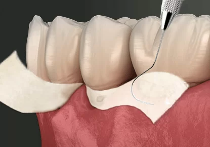
  • CONNECTIVE-TISSUE GRAFTS
  • FREE GINGIVAL GRAFTS
  • PEDICLE GRAFTS
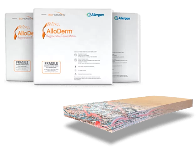
  • ACELLULAR DERMAL MATRIX

Connective-tissue grafts are the most common technique to treat root exposure around one or more teeth. During the procedure, the dentist cut a flap of skin from the roof of the mouth and remove tissue from under the flap. The flap is then stitched back down. The removed tissue will then be stitched to the gum tissue surrounding the exposed root. Over time the attached tissue grows together with the gum tissue forming a higher gum line around the tooth.

Advantages of Connective-tissue grafts: Easier to establish and maintain blood supply, less invasive, more rapid healing, and more natural.

Free gingival grafts are similar to connective tissue grafts. However, in this one, the specialist takes the corrective tissue from the palate to place it in the gum area. Dentists prefer this method for those who have naturally thin gums.

Advantages of Free Gingival Grafts: Prevents a more serious gum recession, creates a healthy barrier of gum tissue, and helps to eliminate the frenum pull.

The pedicle graft method does not take graft tissue from the roof of the mouth. Instead, the gum tissue is grafted from gum tissue around the tooth needing repair. In this method, the flap (called a pedicle) is partially cut away so that one edge remains attached. The dentist pulls up (lower jaw) or down (upper jaw) the flap to cover the root and sew in the place. Dentists prefer this technique for patients who have plenty of gum tissue near the tooth needing repair. In addition to natural grafts from the patient, some dentists prefer to use graft material from a tissue bank.

After the procedure, you will be able to go home. However, if you received a sedative to relax you will need a friend or family member to drive you. Be sure to follow all instructions your doctor gives you for post-operative care. Do not floss or brush the gum line that was treated until the area is healthy. A special mouthwash will be given to you that will help control plaque during this time.

For about a week after the gum grafting It is advisable to follow a diet of soft foods, such as eggs, pasta, gelatin, and yogurt. Pain varies from patient to patient and depends on many variables including pain tolerance and the type of gum graft performed. Your doctor will likely recommend over-the-counter anti-inflammatory medication or, in rare cases, a prescription pain medication. Many patients are able to return to work within a day or two.

Alloderm Graft uses donated medically processed human skin tissue as a source for the graft. The advantage of this procedure is no need for a palatal donor site. This is Great for Dental Tourism as it is Less Invasive but not always the best option for your treatment. You can check these options directly with the Periodontist.

  • Uses of Acellular Dermal Graft material: Root coverage, Soft tissue flap extension over bone graft, Amalgam tattoo correction, and Soft tissue defect repair
  • Advantages of Acellular Dermal Graft material: Reduces the need for palatal autografts or another second surgical site, the ability to treat larger areas in one surgery, and Provides excellent esthetic results.
  • Disadvantages of Acellular Dermal Graft material: Decreases surgical chair-time, Additional cost, Technique sensitive and Longer healing time.

Acellular Dermal Graft is safe, biologically acceptable, and effective. Dental Surgeons use it for the treatment of gingival recession. It is an effective substitute for autologous grafts in the treatment of recession. Acellular Dermal Graft regularly is well tolerated by gingival tissues and has no adverse effects.

Gum Graft Before & Afters

Check out our patients before and afters. Most treatments are provided on two trips of seven days stay in Agadir.

Materials to Gum Graft

AlloDerm®

AlloDerm® is the Best Material available for Gum Grafting Procedures, made by Allergan and Bio Horizon, two very reputable companies in the Dental and Medical Industry.

It is donated tissue that has been processed to remove cells creating a regenerative tissue matrix. This method provides a tissue graft without having to harvest the material from another area in the mouth. Alloderm® Gum Graft in Morocco provides the structure and protein to help your mouth grow its own new tissue.

The Dental Specialist uses collagen to provide scaffolding, stimulate the cells to grow. The protein helps recruit more cells into the graft and helps reshape and remodel the gums. Since its introduction in 1994, over one million grafts have been successfully placed. Donor tissue is extensively screened and tested and then undergoes proprietary procedures to ensure that the Regenerative Tissue is ready for use in patients.

Emdogain®

Emdigain

Emdogain® Gel helps to regenerate the tissue structures that anchor the teeth.

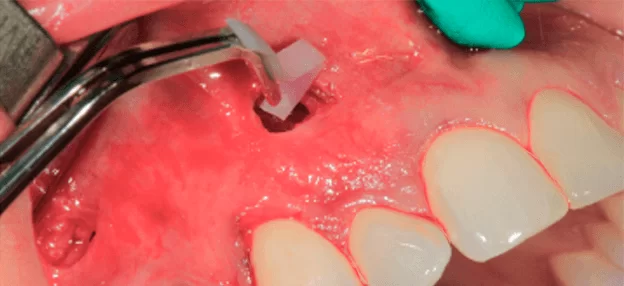
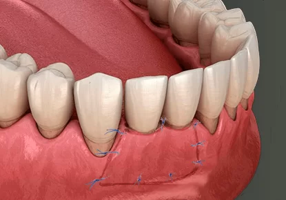
This gel complements Periodontal Surgery, the dentist applies it topically onto exposed root surfaces; It is very effective to stabilize teeth and helps to improve the outcome of periodontal surgery by regenerating the tissue structures that anchor the tooth.

Since its introduction in 1996, Emdogain® has been used in millions of patients worldwide and continues to be a leading product in Periodontal regeneration. The wealth of scientific evidence supporting the product continues to grow. A recent study showed that Emdogain® can increase the predictability of surgical outcomes by achieving equal or better root coverage and attachment. Other recently published studies have indicated that less post-surgical discomfort is reported.Emdogain® Gel is comprised of a number of proteins that self-assemble to create a matrix. The dominant protein in this matrix is amelogenin, the same responsible for tooth development. This protein is present in the human body, therefore, using it doesn’t generate allergic or immunologic reactions. After a single application, it leaves only a resorbable protein matrix on the root surface. No additional surgery is necessary. Regain of clinical attachment and alveolar bone has been proven with the use of

Emdogain
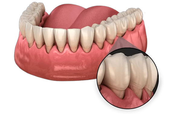
Emdogain® is a biology-based and scientifically proven solution to promote the predictable regeneration of hard and soft tissues lost due to periodontal diseases like intrabony defects, class II mandibular furcation, and recession defects. More than 40 clinical studies, involving 1500 intrabony periodontal defects in 1200 patients, have demonstrated that Emdogain® is effective in stimulating the formation of new periodontal attachment in soft and hard tissue. 60-70% defect fill was measured as a gain of radiographic bone one year following treatment with Emdogain®.

The treatment requires little or no preparation time; no mixing and no specialized products or equipment are necessary. It is convenient and effective to use in areas difficult to treat such as interproximal areas, defects distal to the second molar, defects located under bridgework, and wide defects.

Emdogain Concept

In the biological processes of natural tooth development, Emdogain® forms an insoluble three-dimensional matrix, which allows for the selective colonization of cells. Through cellular interactions, a cascade of events initiates increased cell proliferation, growth factor synthesis, and cell differentiation resulting in the formation of necessary hard and soft tissues such as cementum, periodontal ligament, and alveolar bone.

  1. Attachment – Mesenchymal cells attach to the formed matrix.
  2. Proliferation and growth – The cells spread and populate the surface.
  3. Cementum formation – The cells start to produce cementum by inserting collagen fibers.
  4. Alveolar bone – Along the treated root surface, and at a certain distance, a condensation of fibrous tissue indicates the region where the new alveolar bone is forming.

Types of Gum Recession

Class I

Class I

The recession of the marginal tissue does not extend to the mucogingival junction. The Prognosis for these cases is excellent. Interdental (Between the teeth), there is no Bone loss present and soft tissue is adequate can be narrow or wide. The success rate with these treatments is in the upper 90% and is recommended prior to the gingival recession getting worse.

Class II

Class II

The recession of the Marginal tissue recession extends beyond the mucogingival junction. The Prognosis for these treatments is good. No Mayor boneless and adequate soft tissue can be narrow and wide. The success rate is in the lower 90%. Highly advisable and recommended prior to the gingival recession advancing and creating loss of bone and soft tissue.

Class III

Class III

This type of Gum Recession advanced and extends beyond the mucogingival junction. Tooth structures are highly susceptible to other pathologies, and the treatment does not fully cover the roots. The tooth may be in the wrong position and soft tissue and bone loss are present.

Class IV

Class IV

In this classification, the patient presents teeth severely malpositioned, severe bone loss, and loss of interdentally soft tissue. The recession extends past the mucogingival junction. The Prognosis is Very Poor. Other Treatment options might be available, Severity of treatment is not adequate for dental tourism unless you are willing to fly back to Agadir for multiple trips.

Pinhole Surgical Technique

Pinhole Surgical Technique Perform

To perform a Pinhole Surgical Technique, the dentist uses a pin-like instrument to make a small hole in the gum. This is to reach the tissue and loosen it to cover the retracted area. Then, he places collagen through the hole to keep the gums in place. Most patients are surprised to see an immediate cosmetic improvement. After this treatment, post-operative pain, swelling, or bleeding is minimal. The pin-sized hole typically heals within 24 hours.

What Are the Advantages of Pinhole Surgery?

On a Gum Grafting Procedure, the Dentist makes an incision on the roof of your mouth to remove gum tissue. In some cases, it is necessary to resort to an alternative source or a tissue bank. The Specialist makes another incision and connects the tissue to cover the exposed roots using stitches.

Gum Graft Before Coming to Agadir Pre-Ops

Inform yourself on steps to take for a successful outcome, please read the full instructions, Agadir Dental Specialists thanks you in advance for being an informed patient.

Before surgery (three weeks)

Smoking and dental implants don’t go together. Smoking Is The Main Factor For Dental Implant Failure? We know smoking can be difficult to quit but we insist that patients stop smoking at least three weeks before their dental implants. On unusual occasions, we would ask you to stop smoking sooner, because it can interfere with your recovery, by reducing blood flow, which may prevent the incision from healing. We Highly Recommend You Quit Smoking Once You Commit To Your Dental Treatment

Before surgery (three weeks)

We know you will be on vacation and the all-inclusive bar might be tempting. You must stay away from any alcoholic beverage for a minimum of five days before your procedure. Alcohol causes increased bleeding, and the chances of infection, as well as depress immune functions and might interfere with wound recovery.

Before surgery (three weeks)

Please check with your physician about suspending any medication prior to arrival. We Have No Knowledge Of Your Medical History Prior To Your Arrival To Cancun It Is Very Important You Contact Once Of Our Dental Coordinator And Let Them Know What Medicines You Are Taking. Sometimes We Will Ask For A Letter From Your Physician To Suspend Certain Medication During Procedures Or Healing Process

Gum Tissue Graft Recovery

Do NOT eat anything until the anesthesia wears off, as you might bite your lips, cheek, or tongue and cause damage. You will probably have some discomfort when the anesthesia wears off; take correctly your non-aspirin pain medication(s).

Informed patients are happy patients that why we have taken the time to write this.
Any questions please give us a call at 1 888 231 8041

Gum Graft Complications or Failure

Complications of a Gum Graft are rare. Infections are uncommon. But see your doctor right away if you notice any oozing or unexpected bleeding around the Graft site. In rare cases, the Graft tissue may not properly adapt to the graft site. You may need to have the procedure performed again if this happens. If you do not like the appearance of the graft after healing time, talk with your periodontist about gingivoplasty (reshaping of the gum tissue). They may be able to modify the tissue to make it look more pleasing to you.

  • Severe swelling/loosening sutures
  • Tissue sloughing from upper arch
  • Infection
  • Uneven healing

The survival of the graft tissue (generally obtained from the roof of the mouth) depends on the blood supply. That comes from the surrounding bone (since there are no blood vessels on tooth surfaces). The stability of the grafted tissue is very important for the newly forming and migrating vessels.

Severe swelling and/or loosening of the sutures during healing can cause surgery will not be successful. In such cases, it is necessary to wait until the maturation of the surrounding tissues is complete and try again. This does not happen very often. However, every case is different and some surgical sites are harder to deal with. It will also influence the severity of the exposure and quantity of soft and hard tissue support.

A recent study showed that it can increase the predictability of surgical outcomes. Endogain achieves equal or better root coverage and attachment.

However, the Experts in Oral Health suggest the fabrication of a plastic vacuum form of your upper arch. This snaps press-fit onto teeth to make sure that the palate is covered in its entirety. Thus, it provides pressure to the donor site after the surgery. Also, it avoids irritation from food and the tongue. If you are expecting a series of grafting surgeries, this is a good option.

Similar to any type of surgical procedure, infection will delay wound healing. This is especially true for oral wounds that are exposed to continuous bacterial challenges. A prescription for antibacterial/ antiseptic mouth rinses is generally given to control bacterial load for the first 10 days of healing.

Following complete wound healing, if there is an obvious problem with symmetry, it may be necessary to go back and do what is called “gingivoplasty”. This is a simple procedure performed to thin the extra soft tissue.

Gum Graft FAQs

Gum Graft is a specific alternative to treat gum recession. There are different types of Gum Graft but, in principle, all tend to restore the gum lines to healthy levels. Complementary and preventive procedures are Scaling and Root Planning (Periodontal Cleaning).

Periodontal disease is the main cause of gum recession along with other factors like poor oral hygiene and genetic predisposition; also other causes involve smoking, improper brushing, and illnesses such as diabetes and hormone disorders.

The most common is Connective-tissue Graft which involves the surgical removal of some portion of tissue from the roof of your mouth; However, at CDS we can perform any of the techniques presented on this page.

After the Specialist identifies the amount of tissue that he will remove and place later in the addressed area. Then the dentist administers local anesthesia and proceeds to make the incisions. The Specialist uses a Water Lase machine to finish the procedures faster and more accurately. Also, allows the patients to expedite recovery after surgery. Water Lase reduces the risk of infection and allows the specialists to remove more tissue per session. After that, the tissue is placed and sutured in the addressed area.

Unfortunately, no! Long exposed roots, notches in the teeth (called abfraction notching), roots that have fillings, and teeth that have lost bone between the teeth are difficult to predictably cover. Nonetheless, almost always, they can be improved! The photo to the right shows all of these problems. This underscores how important it is to have treatment before things get too severe.

It is important to realize that as the gums recede, the bone around the teeth dissolves. Thus, in adults, all gum recession is also bone loss! The bone of course is what holds the teeth in place. Don’t delay! Keep your teeth healthy and beautiful!

Yes. Covering the exposed root will decrease the sensitivity. There are some teeth that are sensitive but may not need gum grafting to improve the sensitivity. We will show you how.

After surgery, most possible swelling, bleeding and soreness disappear within the first week after the procedure is done. However, the complete time frame of healing is of 4 weeks, which involves certain medication and the avoidance of certain foods for the time indicated by the specialists.

Treatment outcome is generally evaluated 4-6 weeks after soft tissue grafting. You need to wait up to 8 weeks or so for tissue maturation and to determine how much tissue has “taken”. If the graft tissue was thick to start with, the superficial layer will slough off but the deep parts will survive. (The most outside portion of the graft would not survive due to lack of blood supply. It becomes necrotic-white and comes out.) What is left should be reddish in color and will survive. White/pink color may change based on tissue thickness.
Our specialists will need to re-evaluate the area and determine how much of it has taken. Sometimes it may be necessary to do a second surgery (gingival flap surgery) to bring what has “taken” more towards the root surface (coronally).

Yes, the hole will close up. This is just a matter of time. The pain that you will experience is due to the exposition of connective tissue to the oral cavity and by rubbing the tongue and food. 14 days after surgery, the superficial layer of the skin of the donor site will become thicker. The donor site will start feeling much better once this happens. Usually, it takes between 6 to 8 weeks after the surgery for the depression in the tissue to fill up completely.

During the healing, soft tissue shrinks a little, and edema (swelling) goes down. Soft tissue binds the underlying root surface and neighboring bone surface and starts to mature. The most important aspect of this healing is the formation of new vessels that will bring the blood supply into newly grafted soft tissue (angiogenesis). Again, wound stability is very important for the newly-forming blood supply (vascularization).

Soft tissue healing generally takes 4-8 weeks to return to normal. Healing time also depends on how much tissue (thickness and area) was taken from the roof of the mouth and whether you had any bone exposure after the procedure. If you had bone exposure at the donor site, the healing may take longer. Also, if you developed bleeding into the tissue which accumulated there and caused swelling (hematoma together with hemorrhage), this may delay the wound healing.

Given the right indication and right surgical technique, gum graft surgery works very well most of the time. Factors that help to predict the success of the procedure are the type of defect that needs treatment and the amount of stress put on the graft after surgery.

The type of defect is important for graft survival. Narrow defects with intact tissue between the teeth (interdental areas) have a greater chance of success since the root surface does not have any blood supply and adjacent tissue will help the graft tissue to survive. Complete root coverage is the predictable outcome when this type of defect is present. The success rate of root coverage decreases with increasing interdental bone loss.

Anything that will cause the grafted tissue to move during healing, such as muscle pulls from inside the lip, will affect the healing. That is why periodontists generally show the site to the patient right after surgery and ask him/her not to look at it again during the first week. They know that every time the patient pulls his/her lip to look at it, there is a big chance that the graft will move.

Scroll to Top