3D CBCT X-Ray
What is 3D CBCT X-Ray?
Restore the structure, function, and look of your teeth
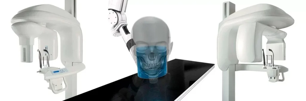
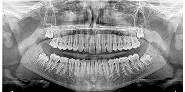
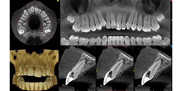
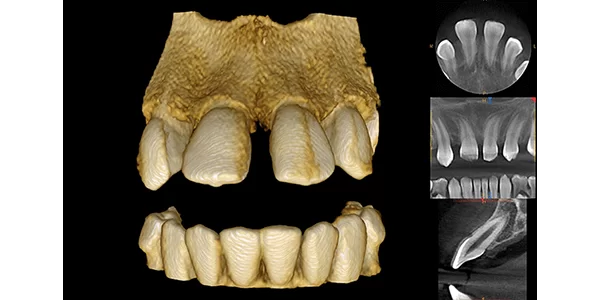
Cone Beam Computed Tomography (CBCT) is a unique type of X-Ray that allows a Dental Surgeon to see the Craniofacial structures, bone, soft tissues, and even nerve pathways of a patient, all in a 3D Image. Unlike Panoramic X-Rays which are 2 dimensional, the CBCT scan allows doctors to see buccal/Lingual distance, bone quality and surgically plan the placement of each implant.
Advantages at a Glance
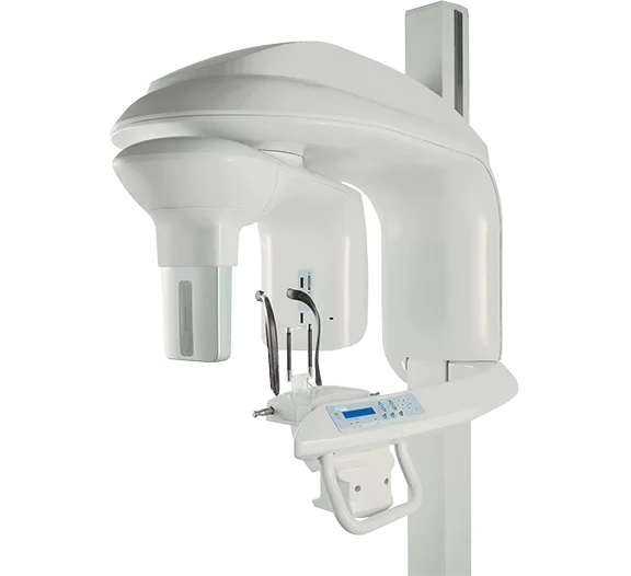
Suitable for a broad range of dental, sinus and temporal bone applications.
Panoramic, CBCT and optional one-shot cephalometric imaging in one solution.
Selectable CBCT fields of view from 5 cm x 5 cm to 17 cm x 13.5 cm.
Very low dose and superb image quality up to 90 μm resolution.
Advantages at a Glance
A 3D CBCT Scan is done as part of your initial evaluation for Implant placement. You should wear loose-fitting clothes and will be asked to remove any metals from your body, including earrings, piercings, hairpins, removable dental appliances, hearing aids, etc.
The 3D CBCT X-Ray is not only useful in the surgical planning of Implants, but it is also helpful in detecting tumors and diagnosing Temporomandibular Joint Disorders (TMJ).